We announced to you in February 2022 the purchase of an EEG system compatible with neurostimulation thanks to a grant from the Natural Sciences and Engineering Research Council of Canada (NSERC). After reorganizing the laboratory and installing our new equipment, we started our first study in 2022, after obtaining approval from the Ethics Committee of the Sectoral Research in Neuroscience and Mental Health (CIUSSSCN; project #2022-2478).
Today, we present to you this exciting project aimed at better understanding the effects of neurostimulation on brain activity in individuals aged 20 to 85 years old in order to optimize the use of neurostimulation and induce as many benefits as possible. Indeed, it is well known that some individuals respond to stimulation, demonstrating beneficial effects, while others do not. However, the factors underlying this significant variability are still unknown. Age could be a factor, as well as pre-stimulation brain activity. For example, a person with more brain activity in a specific frequency band (e.g., gamma) may be more or less responsive to stimulation. Additionally, the duration of the effects of stimulation on brain activity still needs to be clarified, as well as the extent of the impacts. These effects have been measured in several studies, but not in the regions that interest us in the laboratory, namely the regions that control speech and language.
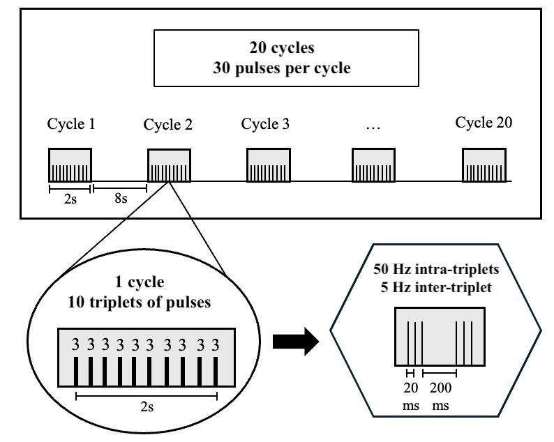
Figure 1. Intermittent Theta-Burst Stimulation (iTBS). 600 pulses are delivered over 20 cycles of 10 triplets of pulses in three minutes. One cycle lasts 10 seconds, and the total duration is 200 minutes. Used with permission from Valérie Brisson 2024 (adapted from her doctoral thesis).
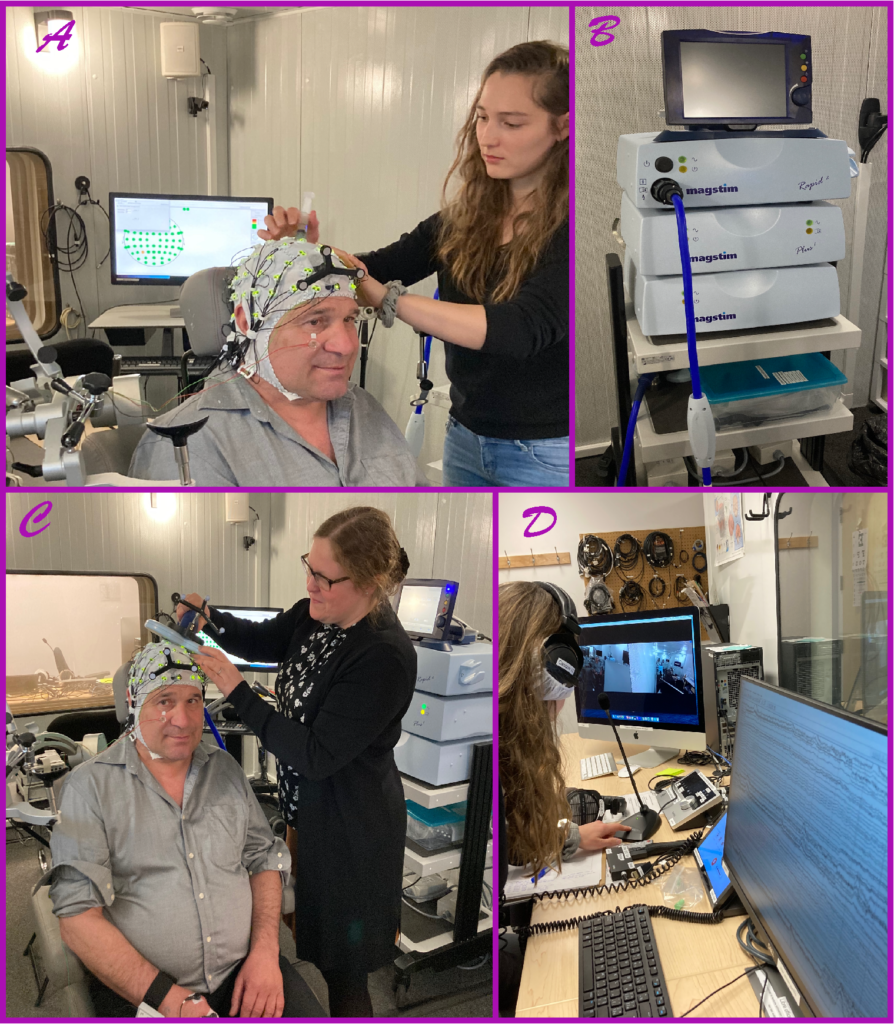
We have recently used this protocol in two studies conducted by Valérie and Pascale to improve speech perception in noise. The results are promising but modest. TMS is a technique that allows painless stimulation of the human brain by rapidly alternating a magnetic field induced in a coil of copper wire placed on the head (Figure 2B and C). The magnetic field is generated by a current that flows through an insulated coil of wire in a plastic sheath. The current generates a magnetic field, which in turn induces a secondary current in a very small part of the brain. The effect of TMS is to modulate (amplify or decrease) the activity of a brain area (a few square millimeters) temporarily (from a few seconds to a few minutes).
In the project, we target three areas involved in speech processing. These regions are identified using brain images of each individual obtained by magnetic resonance imaging in our brand-new MRI platform at CERVO. This allows us to consider individual anatomical characteristics (brains are all different!). MRI is an imaging technique that provides high-quality images of the body and brain. These images are generated using a magnetic field and radio frequencies produced by the MRI machine.
During the project, brain activity is measured using electroencephalography (EEG), before and after stimulation. EEG is a non-invasive and safe technique that measures the electrical activity generated by neurons using small electrodes placed on the head (Figure 2A and D). The electrical activity of the brain is measured before and for 60 minutes after stimulation to determine when it returns to baseline. This 60-minute period is divided into several short intervals. During recordings, participants must remain still and at rest. This allows us to study the baseline brain activity, which is considered an indicator of brain health.
Figure 2. Example of a data collection session within the context of the project. A. Jessica prepares a participant by applying gel for the electrodes. When all the electrodes are filled and their green light is on, the participant is ready! B. The TMS device. C. Pascale holds the TMS coil and applies brain stimulation. D. Jessica monitors brain activity recordings outside the lab's soundproof room. Our new cameras allow us to ensure the participant's well-being, and we can communicate with those inside the room using a system of microphones and speakers.
Our objective is to recruit 25 healthy individuals with no contraindications to MRI and TMS. The study is quite complex, comprising four visits: the first visit takes place at CERVO’s MRI imaging center to obtain participants’ brain images, and the other three take place in the soundproof room of our laboratory. During each session at the lab, a different brain region is stimulated with TMS. This will allow us to compare the effects of stimulation on different language-related brain areas. So, approximately 100 data collection sessions are expected, nearly 10 hours per participant! Furthermore, since the study combines MRI and TMS, recruitment is challenging due to the numerous exclusion criteria aimed at ensuring participant safety.
Led by Pascale, our lab director, the study has enabled us to train several students, which is one of the main objectives of our grant: Juan, Camille, Marianne, Thomas, Keir, Marie, and Jessica. We are expecting a new intern from France, Marion, who will join the project starting in May. The equipment is now being used in a second project in the lab (NeuroSPiN) and will soon be used in a third project, thereby increasing training opportunities.
Although we do not yet have results, we have now completed data collection for 13 participants and will begin data analysis this summer. We will also collaborate with Professor Philippe Albouy for EEG data analysis. Our goal is to complete data collection by the end of summer or early fall at the latest. A big thank you to all who have participated in the project and contributed to data collection and project setup!
Stay tuned for our first preliminary results at the end of the summer!