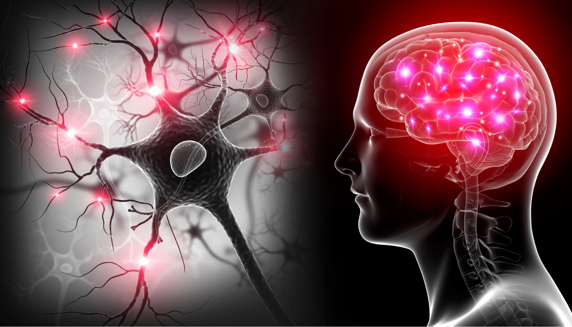
Photo credit: Brain and nerve cells, illustration by Science photo library on Canva.
Neurons are microscopic cells, of which we have many: it is estimated that the human brain contains 100 billion neurons (Purves et al., 2015)! Neurons communicate with each other by transmitting electrical and chemical signals. It is thanks to their morphology and their complex organization that neurons can communicate, which is essential to the functioning of the human brain.
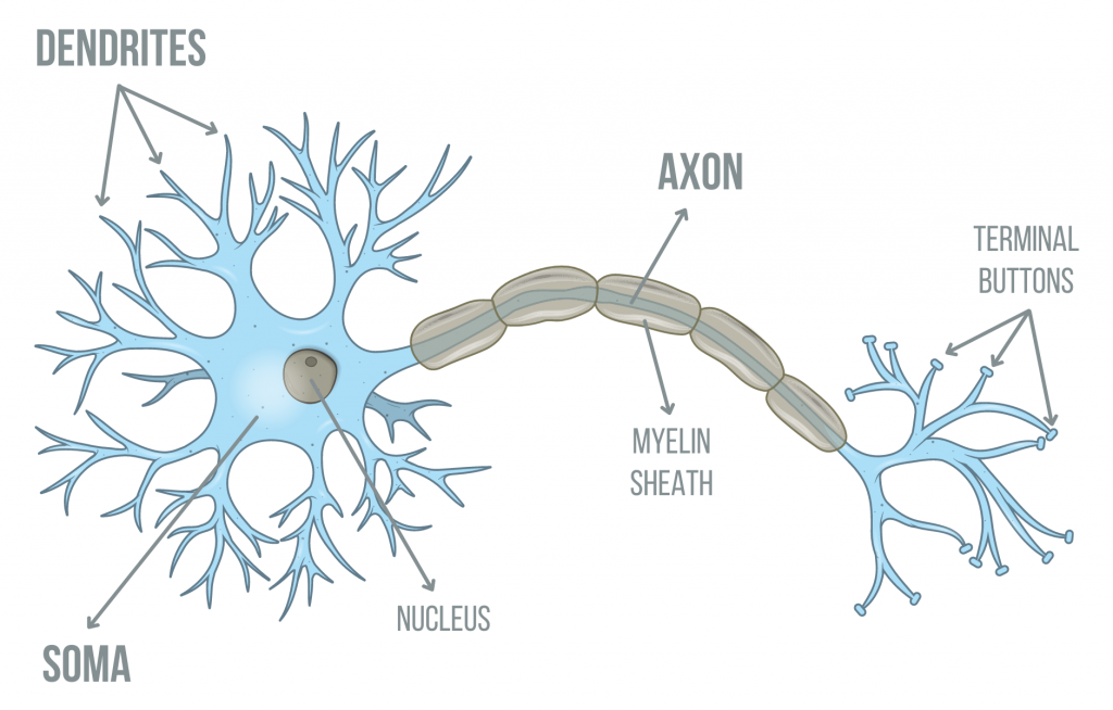
Neurons are composed of three parts: the soma (or body), the axon, and the dendrites (Bear et al., 2007; see figure 1).
Figure 1. Diagram illustrating a neuron and its principal components.
The soma is also called the cell body of the neuron. It contains many structures which are surrounded by membranes, including the nucleus (the other structures of the cell body are not illustrated in figure 1 and will not be discussed in this post). The nucleus contains the genetic material (DNA). Reading the DNA’s genetic code allows for the synthesis of proteins in the soma. These neuronal proteins are destined to numerous functions. Among other things, they are inserted in the neuronal membrane (which separates the neuron from its environment) and form certain passageways for molecules. Some of these molecules, which we call “neurotransmitters”, are transmitted from one neuron to another. Neuronal proteins thus contribute to the transduction of the nervous impulse, which is generated in the soma (Bear et al., 2007).
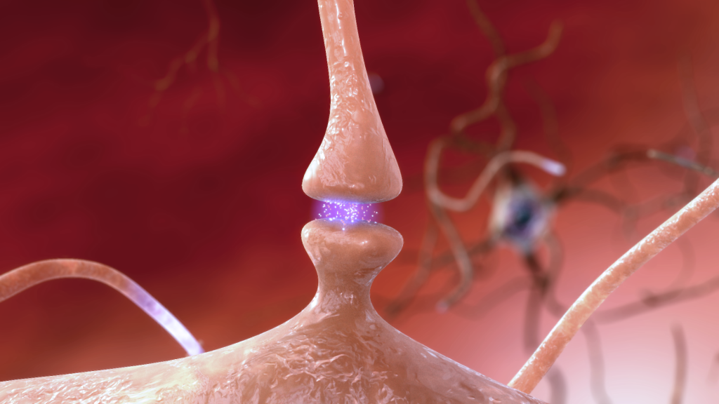
The axon is a structure whose length can vary from less than a millimeter to more than a meter, depending on the type of neuron (Bear et al., 2007). The axon is covered by an isolating membrane, the myelin sheath, which helps speed up the propagation of the electrical impulsions (Bear et al., 2007). The axon is involved in the transport of nervous impulses. The electrical impulses propagate in the axon from the soma of the neuron to the terminal buttons, located at the end of the axon (they are also called axon terminals). At the terminal buttons, the neuron is in contact with other neurons. This area of contact is called the synapse (see figure 2). In the majority of cases, as illustrated in figure 2, there is no direct contact between neurons. When an electrical impulse reaches the terminal buttons, neurotransmitters are released in the space between neurons, called synaptic cleft, then are captured by the receiving neuron. The nervous impulse is thus propagated from one neuron to another via electrical and chemical signaling.
Dendrites are extensions of the soma and look like the branches of a tree; the word dendrite means tree in Greek (Bear et al., 2007). The dendrites are where the neuron receives information from other neurons. Dendrites are covered in receptors that capture the neurotransmitters released by other neurons at their terminal buttons. One single neuron can receive information from many other neurons – up to approximately 100 000 (Purves et al., 2015)!
To summarize, when a neuron captures neurotransmitters at its dendrites, an electrical impulse can be generated in its soma; this impulse propagates down the axon to the terminal buttons where neurotransmitters are released. This series of events occurs from one neuron to the next.
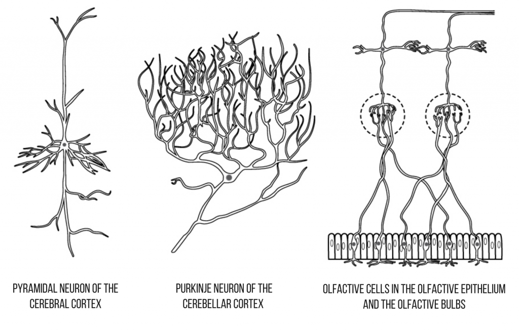
There exist different types of neurons, which are classified according to the shape of their dendrites, the length of their axon, or their function. For example, there are pyramidal neurons, Purkinje neurons, and olfactive neurons, three neuron shapes that are quite different from one another (figure 3).
It is important to mention that neurons are not the only cells in the nervous system. The nervous system also contains glial cells, which are approximately three times more abundant than neurons and play many essential roles. The glial cells influence the establishment and maintenance of synapses and modulate the transmission of information between neurons. Certain types of glial cells (oligodendrocytes and Schwann cells) make the myelin that wrap the neurons’ axons (Purves et al., 2015). Certain glial cells play a role in the inflammation process in the central nervous system, a mechanism involved in many neurodegenerative diseases.
In the field of cognitive neurosciences, it is not possible to observe or study the functioning of individual neurons in alive individuals because there are no tools available. However, we can measure the simultaneous electrical activity of thousands of neurons by using electroencephalography (EEG) or induce the transmission of electrical impulses within brain networks (large collections of neurons) using transcranial magnetic stimulation (TMS). With magnetic resonance imaging (MRI), it is possible to visualize the gray matter, which contains the neurons’ cell bodies, as well as the white matter, which is mainly composed of axons, even though we cannot see the individual neurons.
In a future blog post, you will learn more on the synaptic transmission and neurotransmitters, that is, on the way the nervous impulses are transmitted from one neuron to another at the synapse. Stay tuned!
Here are the works that were used to write this blog article and which you may consult to learn more about neurons:
Purves, D., Augustine, G.J., Fitzpatrick, D., Hall, W.C., LaMantia, A.S., & White, L.E. (Eds). (2015). Neurosciences (5th ed.). Sinauer Associates.
Bear, M.F., Connors, B.W., & Paradiso, M.A. (2007). Neurosciences: Exploring the brain (3rd ed.). Lippincott Williams & Wilkins Publishers.

