Photo credit: Man hand brain by Aramyan on Canva.

First of three texts in our series on the human brain.

What is the brain made up of? What are its structures?

In this blog post, we invite you to discover the brain, one of the most important organs of the human body.

The brain is part of a system of organs called the nervous system. This system is made up of two subsystems, the central nervous system (CNS) and the peripheral nervous system (PNS). In this post, we will discuss the CNS, which includes the brain. The CNS consists of two main structures, the encephalon and the spinal cord (see figure 1).

Figure 1. Diagram of the central nervous system in humans (Human Brain Illustration) adapted from the original image by OpenClipart-Vectors on Canva.

The encephalon

The encephalon is made up of 4 main regions, from top to bottom:

1. The brain makes up most of the encephalon. It weighs about 1.35 kg. The brain is divided into two hemispheres – left and right – by a vertical membrane called “ the falx cerebri ”. These hemispheres are connected at the level of the corpus callosum (see figure 1), which allows the hemispheres to exchange information. Each hemisphere is subdivided into 4 lobes: the frontal lobe, the parietal lobe, the temporal lobe and the occipital lobe (Figure 2). These lobes are named after the bone of the skull under which they are located (for example, the frontal lobe is located under the frontal bone). There is a fifth invisible subdivision of the surface, the insular lobe. This lobe is located inside the brain and is covered by the parietal, frontal and temporal lobes.

Figure 2. Diagram of the lobes of the brain and the bones of the skull.

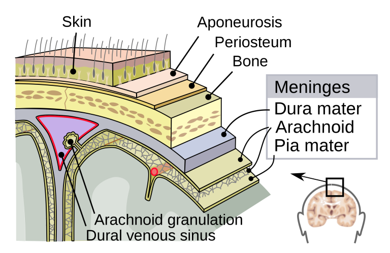
The brain is involved in a very large number of functions, including the processing of sensory information from sensory receptors (e.g., eyes, ears, etc.) as well as the production of voluntary movements. It is also the center of personality, intelligence, consciousness and cognitive functions.

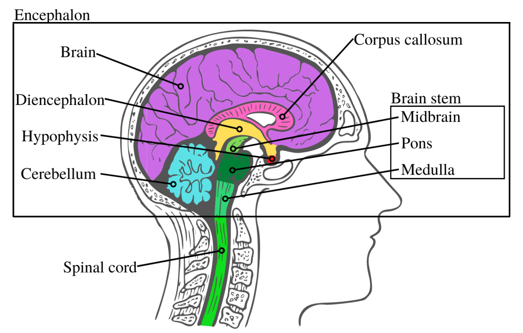
2. Another part of the encephalon is the diencephalon, which is located inside the brain, above the brainstem (see figure 1). The diencephalon comprises three sub-regions:

a) The thalamus, which measures approximately 3 cm and corresponds to 80% of the diencephalon. It is made up of two twin regions of oval-shaped gray matter.

b) The hypothalamus, located under the thalamus, is made up of 12 nuclei distributed in 4 regions (mammillary, tuberal, supraoptic and preoptic), each involved in specific functions.

c) The epithalamus is located above and behind the thalamus. It is composed of the pea-sized pineal gland, the habenular nuclei and the medullary streak.

The diencephalon is involved in the transmission of sensory information to the cerebral cortex (upper layer of the brain which contains billions of neurons), as well as in the planning and regulation of voluntary movements, emotions, memory and cognition and the secretion of hormones, including melatonin.

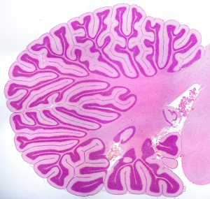
3. The cerebellum, a beautiful tree-like structure (see Figure 3), is located at the back of the brainstem, below the brain. Although its mass is only 10% of the mass of the brain, it contains almost half of all neurons of the encephalon.

Figure 3. Illustration of the cerebellum, seen from the center, staining with hematoxylin and eosin, photo taken under a brightfield microscope.

Photo credit: Cerebellum by Jose Luis Cavol Martin & Jose Enrique Garcia-Mauriño Muzquiz on Canva.

4. The brainstem is also part of the encephalon. It is the extension of the spinal cord (see figure 1); it is subdivided into three regions:

a) The medulla, located in the lower part of the brain stem and measuring about 3 cm, is the direct extension of the spinal cord.

b) The pons, i located directly above the medulla, just in front of the cerebellum, and which measures about 2.5 cm.

c) The midbrain is located between the pons and the diencephalon, and it is approximately the same size as the pons.

The brainstem serves as a relay for many nerve impulses between the different parts of the CNS. It is also in this region that several involuntary actions of the human body are regulated, such as breathing and heartbeat.

The encephalon contains 4 ventricles (see figure 4) inside of which circulates a liquid called cerebrospinal fluid. The two lateral ventricles are located in each of the hemispheres of the brain. The cerebrospinal fluid is produced within the ventricles. The third ventricle is located above the hypothalamus, between the two thalami (right and left). The fourth ventricle is located between the cerebellum and the brain stem. The lateral ventricles and the third ventricle communicate via small structures called the interventricular foramina; the third and fourth ventricle are connected by the cerebral aqueduct.

Figure 4. Illustration of the ventricles of the brain (Ventricles of the Brain) by BruceBlaus licensed CC BY 3.0.

The Spinal cord

The spinal cord essentially plays a role in conveying information to and from the brain, and it plays a major part in producing the spinal reflexes, such as quickly removing your hand from a hot object. Two types of pathways run through the spinal cord: the sensory pathways, which are ascending, conveying nerve impulses from the sensory organs to the brain, and the motor pathways, which are descending, conveying nervous impulse from the brain to the body, allowing us to move. The spinal cord is connected to the body by 31 pairs of nerves called spinal nerves (as opposed to the 12 pairs of cranial nerves that emerge from the brain) which branch out to form a nerve tree (Figure 5).

Facebook_Schéma_anatomie_SNC_Fig5_ENG

Figure 5. Illustration of spinal and cranial nerves
 Nervous System Icon par Becris on Canva

CNS structures are highly protected. Several structures protect them:

  1. Bone tissue includes the 8 bones of the skull that protect the brain and the 26 vertebrae that protect the spinal cord.
  2. The 3 layers of meninges, called dura mater, arachnoid, and pia mater (see figure 6) lie between the bone tissue and the brain or spinal cord.
  3. The cerebrospinal fluid (CSF) circulates in the four ventricles of the brain, but also in the subarachnoid cavity and in the central canal of the spinal cord. The CSF has three main functions:

a) Mechanical protection: the CSF protects the brain and spinal cord from jolts and shocks.

b) Chemical protection: the CSF allows the proper transmission of nerve impulses in the CNS by regulating its chemical environment.

c) Circulation: the CSF allows the exchange of nutrients and waste products between the blood and the nervous tissue.

Figure 6. Illustration of the protective layers of the CNS (Meninges-en) by (Jmarchn) licensed CC BY-SA 3.0

That’s it, it’s already the end! This post has provided an overview of the main components of the central nervous system. As the brain is a highly complex organ, it is impossible to describe it in one post. In the next publications of this series, we will discuss the gray and white matter, as well as the functional organization of the brain, in particular the cerebral cortex.

Reference consulted to write this blog post:

Tortora, G. J., & Derrickson, B. (2007). Principes d’anatomie et de physiologie (M. Forest & L. Martin, Trans. 2e ed.) : ERPI.

Further readings:

, ,