Third of three texts in our series on the human brain.
 
In the first two posts of this series, we discussed the anatomy of the human brain, and more broadly, the anatomy of the central nervous system (CNS). In this third and final post, we discuss the functional organization of the brain, that is, how the brain accomplishes various functions such as processing information from sensory receptors (e.g., ears, eyes , etc.), talking, or controlling body movements.

Much of the brain’s functions are governed by the cerebral cortex. During its development, the cortex forms folds that are called “gyrus” (or gyri in the plural). These gyri are separated by grooves of various depths. The deepest ones are called “fissures” while the shallower ones are called “sulcus”. If unfolded completely, the cerebral cortex would occupy an area of ​​about 2600 cm2, or about a square the size of a chessboard!

The cortex is divided into dozens of regions (or areas) based on their functions. These regions can be grouped into three main categories: sensory, motor and associative. To facilitate the identification and location of areas, a numbering system was developed more than 100 years ago, which is still used today. This map of the cortex, shown in Figure 1, bears the name of Brodmann’s areas, the name of its creator. Regions are numbered based on their internal structure. Indeed, regions with different types of functions (sensory, motor and associative) have different cellular architecture. That is, they contain different types of neurons, and their connectivity is also different. The areas described in this post will be accompanied by their numbering in parentheses and will refer to Brodmann areas.

Figure 1. Illustration of Brodmann’s areas of the lateral and medial view of the left hemisphere. Adapted from Brodmann areas by Vysha under licence CC0 1.0.

Sensory Areas

Sensory areas, as their name suggests, receive sensory nerve impulses from sensory receptors in the body (eyes, ears, nose, fingers), and play a role in sensation and perception. Sensory information is conveyed by the ascending pathways, relayed by the thalamus (except for smell) and transmitted to the “primary” sensory areas. There are two types of sensory areas, primary areas and secondary areas. The primary sensory areas are the gateways for sensory information into the brain. The primary areas carry out a first level of processing of sensory information, while the secondary sensory areas carry out a second, finer level of processing. There are several primary sensory areas:

1. The primary somatosensory area (areas 1, 2 and 3). This area is located behind the central sulcus of each cerebral hemisphere, in the parietal lobe (Figure 1). This area receives nerve impulses from several types of sensory receptors, including touch receptors (mechanoreceptors), pain receptors (nociceptors) and temperature receptors (thermoreceptors) located throughout the body. Each region of the body is represented in the primary somatosensory area. The size of this representation is not proportional to the size of each part of the body. Instead, it is proportional to the number of sensory receptors that each region of the body contains. For example, the fingers and the mouth are small, but because they contain many receptors (and are therefore highly sensitive), they occupy a larger cortical surface than the hips, which are larger, but contain few receptors (sensations originating from the hips are thus less precise). The primary somatosensory area therefore contains a map of the body (often called a “homunculus”) that allows it to identify the origin of a stimulus. A representation of this map is shown in Figure 2. A lesion in the primary somatosensory area leads to an alteration or loss of sensations related to touch or pain either globally or only in the body part representation that is damaged.

Figure 2. Illustration of sensory homunculus (1421 Sensory Homunculus) by OpenStax College under licence CC BY 3.0.

2. The primary visual area (area 17). This area is located at the posterior end of the occipital lobe and receives information from the eyes. It contributes to identifying the shape and colour of the objects around us. Thus, a lesion in this area inevitably leads to loss of vision in a portion of the visual field.

3. The primary auditory area (area 41).  The primary auditory area is located in the upper part of the temporal lobe, in a region called the transverse temporal gyrus or Heschl’s gyrus (Figure 3). It receives all the auditory information from the ears, including speech, and helps determine the intensity and pitch of a sound (low or high). It is also involved in processing the temporal structure of sounds, such as their rhythm. A lesion in the primary auditory area can lead to deafness.

Figure 3. 3D illustration of the brain and Heschl’s gyrus (Transverse temporal gyrus) by Daniel Sabinasz under licence CC BY-SA 4.0.

4. The primary gustatory area (area 43). This area is located just below the primary somatosensory area, in the parietal lobe. It receives its information from the taste buds of the tongue and thus contributes to the perception of gustatory sensations (taste). In addition to the tongue, the gustatory area receives information from the larynx, mainly from the epiglottis. The larynx receptors do not play a role in taste, but rather in the detection of certain chemical substances and the passage of water, in connection with the role of the larynx in the protection of the respiratory tract during feeding. In addition to taste dysfunction, damage to the gustatory cortex is usually associated with other neurological problems, such as Lyme disease.

5. The primary olfactory area (area 28). Unlike other primary sensory areas, the primary olfactory area is not located on the surface of the cortex, but inside it, near the amygdala and the parahippocampal gyrus. It receives the olfactory information captured by the nose and contributes to the perception of odours. A lesion in the primary olfactory area can lead to a loss of smell. Some diseases can also attack the olfactory system. For example, the SARS-CoV-2 (COVID-19) virus attacks a protein essential to the integrity and proper functioning of sensory neurons in the nose, causing the loss of smell.

Motor Areas

The motor areas are involved in the preparation, initiation, and execution of movements. Unlike sensory areas, motor commands come from the anterior (frontal) part of the cortex. Similar to the organization of the sensory cortex, the motor cortex can be divided into a primary motor area and secondary motor areas. The primary motor area is primarily responsible for controlling the strength and duration of muscle contractions. It is responsible for sending commands to the muscles for the execution of movements. The premotor areas are involved in the integration of cognitive and sensory information. This integration allows these areas to plan actions, especially those that are guided by the senses, such as the ability to grasp an object and lift it. This section will focus on the main motor areas.

1. The primary motor area (area 4). This area, involved in the voluntary and precise contractions of the muscles, is located in the frontal lobe, in front of the central sulcus. It is therefore adjacent to the primary somatosensory area and shares several similarities with this area. Thus, each muscle in the body is controlled by a distinct region of the primary motor area. The size of the region that controls a muscle is not proportional to its size, but to its precision. For example, the region that controls the small finger muscles—which must perform precise and complex movements—is larger than the region that controls the large leg muscles, because leg movements are less precise and less complex.

2. The premotor cortex (area 6). The premotor cortex is adjacent to the primary motor area, with which it communicates. The premotor cortex can be represented as a mosaic of areas. The functions of these regions are usually described separately for the lateral (side) and medial (middle) portions. The lateral and medial premotor areas are thought to be involved in the selection of a movement. However, the lateral premotor area is specialized in initiating movements based on external cues (e.g., in response to an event occurring in the environment), while the medial premotor areas prepare movements based on cues internal (e.g., related to the desire to move from point A to point B).

3. The frontal oculomotor area (area 8). Located in the frontal lobe between the premotor cortex and the prefrontal cortex, the oculomotor area is involved in voluntary eye movements. It is additionally responsible for the sweeping movements of the eyes.

Association Areas

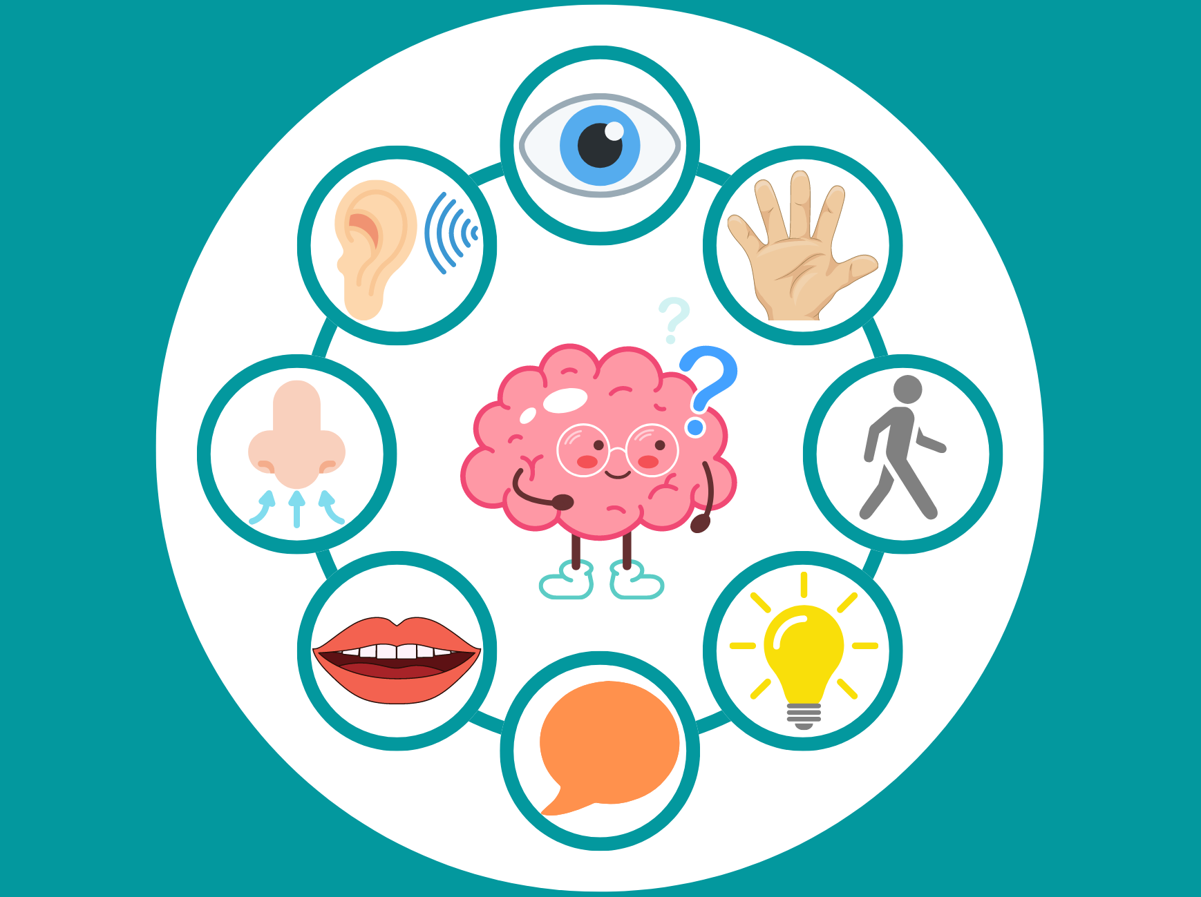
There are two types of associative areas, i.e., unimodal and multimodal associative areas. Unimodal associative areas make it possible to integrate different information related to the same sensory modality (e.g., different visual characteristics such as colour, shape, and movement). They include the secondary sensory areas. A sensory modality, for example vision, can be associated with several unimodal associative areas. Multimodal associative areas are large cortical areas involved in the integration of information from several modalities. They also participate in more complex functions, which are sometimes said to be of a “higher level” than the sensory and motor areas, including memory, emotions, judgment, intelligence and language. Here we describe some of these areas:

1. Visual associative areas (areas 18 and 19). The associative visual areas are located in the occipital lobe, next to the primary visual area. There are four of them: V2 to V5 (V1 being the primary visual area). The division of associative visual areas, and visual areas more generally, is based on the function and structure of these areas. V2 receives information from the primary visual area and would respond, among other things, to colour differences, patterns of medium complexity and the orientation of an object. The information processed at the level of V2 is then sent to the areas V3, V4 and V5, which are specialized in the finer processing of different aspects of the visual signal such as the ability to recognize an object.

2. The associative auditory cortex (areas 22 and 42). The associative auditory cortex, also called the secondary auditory cortex, includes the association areas related to hearing. As for the visual areas, the auditory areas have several subdivisions. The first region receiving auditory information is the primary auditory area, while the others are the associative auditory areas. The associative auditory areas are located below and behind the primary auditory area. They are involved in the phonological processing of speech sounds, including their identification (e.g., distinguishing the /p/ sound from the /b/ sound).

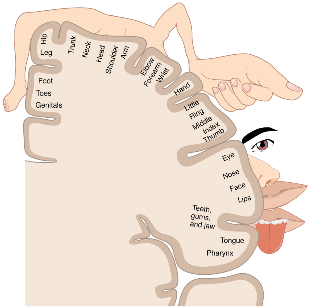
3. The somatosensory associative area (areas 5 and 7). Located directly behind the primary somatosensory area, the associative somatosensory areas receive information from the primary somatosensory area, but also from the thalamus and other regions of the brain. While the primary somatosensory area makes it possible to capture raw information, for example the presence of an object between two fingers, the associative area is involved in the identification of this object through spatial and tactile memory containing past sensory experiences. The somatosensory associative area is linked to the amygdala as well as the hippocampus, a complex structure associated with memory consolidation and decision-making. These connections allow for the integration, at the level of the associative somatosensory area, of sensory information with stored past experiences. Thus, the associative areas receive, process and integrate sensory information from our environment into our past sensory and emotional experiences.

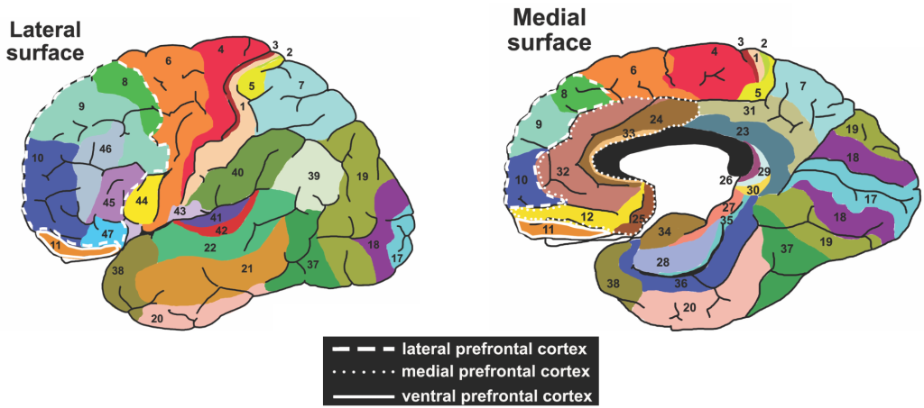
4. The prefrontal cortex (areas 9, 10, 11 and 12). The prefrontal cortex is composed of several areas occupying a large chunk of the frontal lobe, at the level of the forehead. The prefrontal cortex is involved in various functions. It is in communication with several regions of the cortex, as well as with the thalamus and the cerebellum, among others. It is a region that is highly developed in all primates, especially in humans. The prefrontal cortex is the main seat of personality, intelligence, learning, judgment, consciousness, decision-making, mood and many other functions. Lesions to this region can have devastating consequences on these functions.

5. Posterior parietal cortex (areas 5, 7, 39 and 40). This area, also composed of several subregions, is located at the level of the parietal lobe. It receives information from the associative areas, namely the somatosensory area, the visual areas and the auditory areas, but also from two primary sensory areas, the gustatory area and the olfactory area, from the thalamus and even from certain regions of the brainstem. It serves to integrate this information. This multimodal integration is particularly useful for interpreting and reacting to complex multimodal phenomena (such as speech, which is both visual and auditory) and guiding decision-making.

6. Language areas (e.g., areas 6, 8, 22, 40, 44).  Advances in cognitive neuroscience have revealed that language is supported by a very complex network, and that the simplified view that language is supported by two areas (Broca’s and Wernicke’s areas) is no longer adequate. Indeed, areas associated with language are found in all lobes of the cerebral cortex. This includes the inferior frontal gyrus (pars opercularis), the premotor cortex, the supplementary motor area, the prefrontal cortex, the supramarginal gyrus (parietal lobe), the anterior temporal lobe, and the auditory areas. These regions, interconnected by several white matter fasciculi, including the arcuate bundle and the uncinate bundle, are involved in the comprehension and production of spoken or written language.
 
We have now reviewed several areas of the cerebral cortex and their functions. As you can see, the organization of the brain is complex and fascinating!
 
With this post, we conclude our series on the anatomical and functional organization of the human brain. In a future series, we will discuss the neural organization of specific cognitive functions, namely attention, working memory, executive functions, and language. This new series will also address the effect of aging on these functions. Stay tuned!

Reference work used to write this blog post:

Tortora, G. J., & Derrickson, B. (2007). Principes d’anatomie et de physiologie (M. Forest & L. Martin, Trans. 2e ed.): ERPI.

Raju, H. and P. Tadi (2022). Neuroanatomy, Somatosensory Cortex. StatPearls. Treasure Island (FL), StatPearls Publishing

Torrico TJ, Munakomi S. Neuroanatomy, Thalamus. [Updated 2021 Jul 31]. In: StatPearls [Internet]. Treasure Island (FL): StatPearls Publishing; 2022 Jan-.

Obiefuna S, Donohoe C. Neuroanatomy, Nucleus Gustatory. [Updated 2022 Jan 26]. In: StatPearls [Internet]. Treasure Island (FL): StatPearls Publishing; 2022 Jan-.

Donoghue JP, Sanes JN. Motor areas of the cerebral cortex. J Clin Neurophysiol. 1994 Jul;11(4):382-96. PMID: 7962487.

Fogwe LA, Reddy V, Mesfin FB. Neuroanatomy, Hippocampus. [Updated 2022 Jul 18]. In: StatPearls [Internet]. Treasure Island (FL): StatPearls Publishing; 2022 Jan-.

Huff T, Mahabadi N, Tadi P. Neuroanatomy, Visual Cortex. [Updated 2021 Jul 31]. In: StatPearls [Internet]. Treasure Island (FL): StatPearls Publishing; 2022 Jan-.

Purves D, Augustine GJ, Fitzpatrick D, et al., editors. Neuroscience. 2nd edition. Sunderland (MA): Sinauer Associates; 2001. The Auditory Cortex.

Hirano S, Naito Y, Kojima H, Honjo I, Inoue M, Shoji K, Tateya I, Fujiki N, Nishizawa S, Konishi J. Functional differentiation of the auditory association area in prelingually deaf subjects. Auris Nasus Larynx. 2000 Oct;27(4):303-10. doi: 10.1016/s0385-8146(00)00072-9. PMID: 10996488.

Purves D, Augustine GJ, Fitzpatrick D, et al., editors. Neuroscience. 2nd edition. Sunderland (MA): Sinauer Associates; 2001. The Premotor Cortex.

Tremblay P, Dick AS. Broca and Wernicke are dead, or moving past the classic model of language neurobiology. Brain Lang. 2016 Nov;162:60-71. doi: 10.1016/j.bandl.2016.08.004. Epub 2016 Aug 30. PMID: 27584714.

Mountcastle VB. The columnar organization of the neocortex. Brain. 1997 Apr;120 ( Pt 4):701-22. doi: 10.1093/brain/120.4.701. PMID: 9153131.

Tarakad A, Jankovic J. Anosmia and Ageusia in Parkinson’s Disease. Int Rev Neurobiol. 2017;133:541-556. doi: 10.1016/bs.irn.2017.05.028. Epub 2017 Jun 27. PMID: 28802932.

, ,