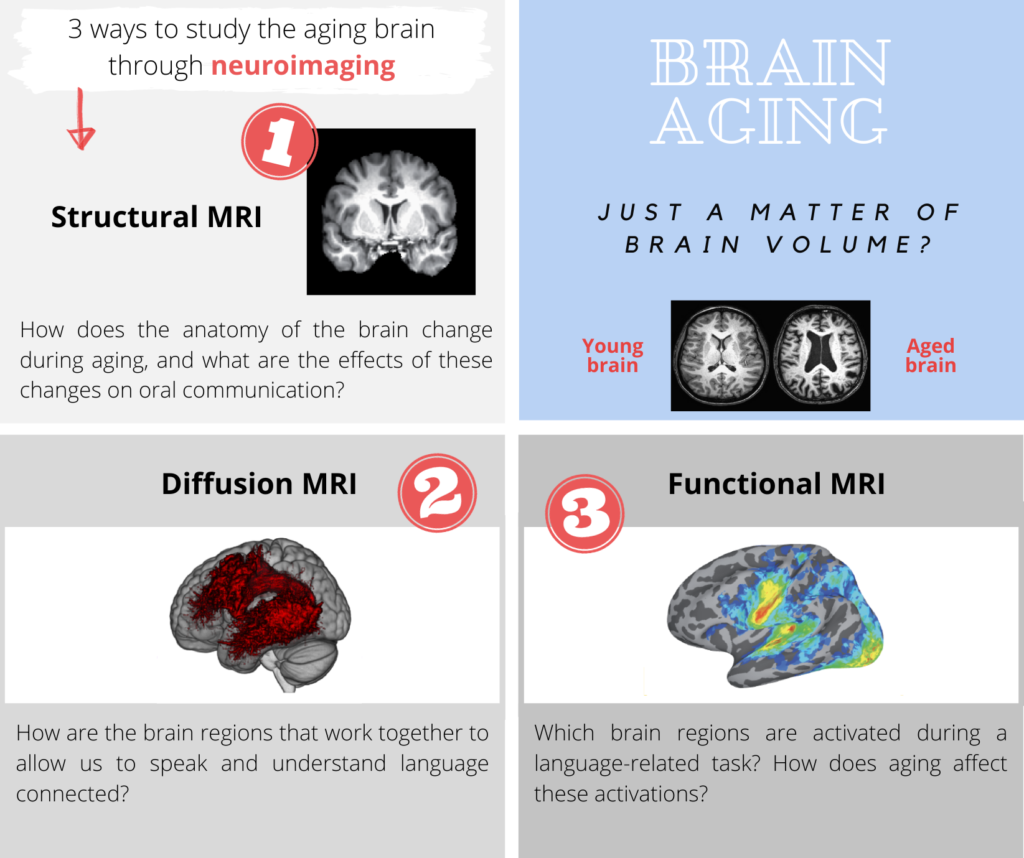
Many of us have seen an image comparing the brain of a young person to that of an elderly person. At a glance, it’s possible to observe the loss experienced by the brain in the course of normal aging. This decrease in brain volume, including gray matter, i.e., the bodies of neurons, is associated with the decline in cognitive and language functions during aging. But is it just a matter of brain volume?
This measurement is certainly important, but it alone does not reflect all the changes in our brain related to age. Indeed, other changes in the structure (see point 2) and functioning of the brain (see point 3) appear during aging. All these changes show that our brain is an extraordinary machine that can change throughout our life, at different rates. These transformations, called plasticity, can affect both the structure and functioning of the brain, and they can be beneficial or detrimental.
Our laboratory is interested in the effect of normal aging on our ability to perceive and produce speech. How can we study brain aging in three ways through neuroimaging?


Structural Magnetic Resonance Imaging (MRI) allows us to obtain images of the brain’s anatomy and measure the surface, thickness, and volume of specific structures such as the insular cortex or the inferior frontal gyrus. Our goal is to identify the neurobiological mechanisms behind communication difficulties that occur during aging. How does the brain change with age? How do these changes impact our ability to communicate? Another interest of our lab involves examining the effect of different types of activities on the aging brain. In a recent project, 192-2017, we found an increase in volume, surface area, or cortical thickness in several brain areas of choral singers compared to non-singers. Some of these differences are associated with our speech perception ability. Furthermore, practicing choral singing may slow down the decline in speech perception. Stay tuned! For more details, please see the list of publications from our lab at the bottom of the page!

This technique allows us to study the organization and health of the white matter, which consists of the extensions of neurons. By studying the movement of water molecules, images of the ‘electric wires’ of our brain can be constructed, and it is possible to extract information about the wear and tear of their microscopic structures or ‘microstructure’. When white matter bundles decline, their ability to transmit neuronal messages is reduced. For example, our lab has demonstrated that there are changes in the microstructure of the arcuate fasciculus in a group of elderly participants, and that this change in white matter is linked to their performance on a speech perception task in noise. The more the microstructure declined, the more performance declined.

This technique allows us to locate brain activity when speaking or listening to someone speak. Is this activation of different intensity between young and elderly participants? Are the same regions activated? Studying language aging is also about understanding how the brain reorganizes itself to compensate for neuronal loss or changes in white matter. These changes can reflect various mechanisms, such as the use of ‘secondary routes’ to continue the work, which may hinder performance, or decreases in activation that could indicate a decrease in power, like an engine gradually losing horsepower. However, decreases in activation can also reflect a form of expertise that allows performing a task with less power. Another type of change involves a compensation mechanism that delays the onset of cognitive declines by keeping or improving performance: the brain works harder or differently to achieve the same performance. Finally, can musical activities such as choral singing or transcranial magnetic stimulation also induce compensatory mechanisms? Several ongoing studies from our lab aim to answer these questions.
These three neuroimaging techniques, combined with behavioral studies and studies involving neurostimulation from our lab, allow us to study normal language aging on multiple fronts. It’s much more than just a matter of brain volume!
References:
Tremblay, P., Perron, M., Deschamps, I., Kennedy-Higgins, D. Houde, J.-C., Dick, A.S., Descoteaux, M. (2018) The role of the arcuate and middle longitudinal fasciculi in speech perception in noise in adulthood. Human Brain Mapping, 40(1), pages 226-241.
– Tremblay, P., Deschamps, I., Baroni, M., Hasson, U. (2016) Neural sensitivity to syllable frequency and mutual information in speech perception and production. NeuroImage, 136(1),106–121.
– Tremblay, P., Deschamps, I., Bédard, O., Tessier, M.-H., Carrier, M., Thibeault, M. (2018) Aging of speech production, from articulatory accuracy to motor timing. Psychology of Aging, 33(7) :1022-1034.


