What makes up gray matter and white matter ? What are the differences between the two ?

Second of three texts in our series on the human brain.

After our post on the anatomy of the central nervous system (CNS), we present in this post the gray matter and the white matter, essential components of the nervous system.

Gray matter and white matter are found throughout the CNS, in the brain, cerebellum and spinal cord. These two substances are not very different at the cellular level. They are both composed of billions of neurons or parts of neurons (it is estimated that the human brain is composed of about 100 billion neurons). However, they have very different roles! The white matter has a role of transporting nerve impulses, and the gray matter has a role in processing the information.

The white matter is essentially composed of myelinated axons, in other words, axons that have a myelin sheath. It is this sheath, which is white, that gives its name to the white matter. The white matter is not organized in the same way throughout the CNS. In the brain and cerebellum (Figure 1B), it is located in the centre, and surrounded by gray matter. On the contrary, at the level of the spinal cord, the white matter is located around the gray matter (Figure 1C).

The length of the axons that compose the white matter differs depending on the function of the neuron. For example, the axons of projection neurons typically range in length from a few millimetres to 1 metre while the axons of neurons that communicate over small distances are shorter, less than a few millimetres.

Facebook_Substance_griseblanche_Fig1_ENG

Figure 1. A) Illustration of the transverse plane (horizontal), in yellow. B) Cross-sectional view of a brain MRI image taken by the Speech and Hearing Neuroscience Laboratory. C) Diagram of a cross-sectional view of the spinal cord (Canva).

In the brain, the axons that make up white matter are bundled together to form nerve-like structures, which are called fibres (the term nerve being reserved for the peripheral nervous system). These fibres connect different areas of the brain. There are several types:

1. Association fibres. These groups of axons transmit information between the different regions of the same cerebral hemisphere. There are 3 types of association fibres. (1) Long association fibres that remain confined to the same region (for example in the motor area) or cross more deeply the white matter of the same hemisphere (such as the arcuate fasciculus). (2) Short association fibres (u-shaped), which connect adjacent regions. (3) Neighbouring association fibres which generally connect different regions of the same lobe (for example in the temporal lobe).

2. Commissural or interhemispheric fibres. The commissural fibres communicate information between the two cerebral hemispheres. For example, the right hemisphere premotor cortex and the left hemisphere premotor cortex communicate through a commissural fiber called the corpus callosum. There are also other interhemispheric fibres, namely the anterior commissure, the posterior commissure, and the fornix (Figure 2).

Figure 2. Illustration of commissural tracts adapted from original image Brain Model by biologycorner under license CC BY-NC 2.0.

3. Projection fibres. These long-range fibres connect very distant brain areas, such as the cortex and the thalamus (thalamic radiation), the cortex and the brainstem and the spinal cord. Projection fibres are categorized into two types: (1) motor tracts, also called descending tracts, which transmit information from the brain to the rest of the body, and (2) sensory tracts, also called ascending tracts, which send the signals picked up by sensory receptors located throughout the body, to the brain. The projection fibres pass through a wide band called the internal capsule (Figure 3), which is located under the corpus callosum. It is important to understand that neurons are unidirectional, that is to say, they can only be involved in one type of tract (up or down).

The gray matter is composed of a greater diversity of neurons parts, but also of other cell types. It includes cell bodies of neurons and their dendrites, unmyelinated axons, nerve endings and glial cells. Glial cells, also called neuroglia, are a collection of cell types that have various functions (mainly to support and protect the neurons) but do not transmit nerve impulses. The glial cells include protoplasmic astrocytes which have a star shape with short extensions, and which would play, among other things, a role in modulating synaptic transmission. They also include microglia and Schwann cells.

The gray colour of the gray matter is due to two factors, the low presence of myelin and the presence of Nissl bodies, structures located mainly in the cell body of neurons, and which have a gray colour.

In the brain and cerebellum, the gray matter is located mainly on the surface and forms the cerebral cortex and the cerebellar cortex respectively (Figure 1B). Gray matter is also found deep in the centre of the brain and form a collection of nuclei. These deep regions of gray matter are called basal ganglia. (Figure 3). The basal ganglia are composed of six nuclei, three in each of the cerebral hemispheres. In each hemisphere, two of the three nuclei are located next to the thalamus, namely the globus pallidus and the putamen. These two nuclei are often grouped together as the lenticular nucleus. The third nucleus is the caudate nucleus. In the shape of a long comma, it “surrounds” the lenticular nucleus (Figure 3). The basal ganglia receive nerve impulses from the cortex and transmit information to the entire cortex through circuits called cortical-subcortical loops. The basal ganglia have many functions. They contribute to the preparation and initiation of voluntary movements (e.g., talking or waving hello), the control of involuntary actions such as swinging the arms when walking or laughing, motor learning, and to the regulation of posture (muscle tone, posture, rigidity). Another important function of the basal ganglia is the regulation of reward and disgust. Finally, but not the least, the basal ganglia are also involved with different cognitive functions, such as attention, selection and inhibition of actions, and consciousness.

Figure 3. Illustration of the basal ganglia, thalamus and amygdala (transverse views on the left and coronal views on the right), adapted from the original image Basal Ganglia Circuits by Mikael Häggström under license CC BY-SA 3.0.

The basal ganglia communicate with several structures in the brain. This includes the thalamus (Figure 3), where all sensory information (except gustatory information), transit before going to other regions of the brain. The thalamus is involved in the perception of pain, temperature, and pressure. The thalamus is also involved in motor functions, allowing communication between subcortical structures (the cerebellum and the basal ganglia) and the cortex, in particular the primary motor and premotor areas, forming what are called cortical-subcortical control loops that we discussed in the previous section. The axons connecting the thalamus and the cortex pass through the internal capsule (Figure 4).

Facebook_Substance_griseblanche_Fig4_ENG

Figure 4. Schematic of connections involving the basal ganglia.

The basal ganglia also communicate with the amygdala (Figure 3), another subcortical structure. The amygdala is involved in the regulation of anxiety, fear, emotional memory, and social cognition (which refers to the cognitive processes involved in social interactions). Information from the amygdala is relayed to the cortex by the thalamus. Another subcortical structure related to the basal ganglia is the substantia nigra (Figure 3), which is made up of gray matter. Along with the basal ganglia, the substantia nigra helps initiate and modulate movement. It is also involved in reward functions (e.g., motivation to perform survival-related actions, such as foraging). Finally, the subthalamic nuclei also interact with the basal ganglia. The subthalamic nuclei are formed of deep gray matter. Along with the basal ganglia, the subthalamic nuclei participate in the regulation of movements.

In the spinal cord, the gray matter is located centrally and is surrounded by white matter. The space occupied by gray matter resembles a butterfly (Figure 1C). The gray matter of the spinal cord has 4 horns, 2 dorsal and 2 ventral horns, which communication between the spinal cord and the body. The nerve impulses carried by the sensory nerves enter the spinal cord at the level of the dorsal horns. Nerve impulses carried from the brain by the motor nerves leave the spinal cord through the ventral horns and convey motor commands to the muscles for the execution of movements.

Now that we’ve discussed the anatomy of the brain, we’re ready to discuss its functional organization, that is, how it is organized to control functions as diverse as walking, grasping, hearing, or talking. The next and last post in this series will focus on the vast topic of the functional organization of the cerebral cortex.

References used to write this post

Tortora, G. J., & Derrickson, B. (2007). Principes d’anatomie et de physiologie (M. Forest & L. Martin, Trans. 2e ed.) : ERPI.

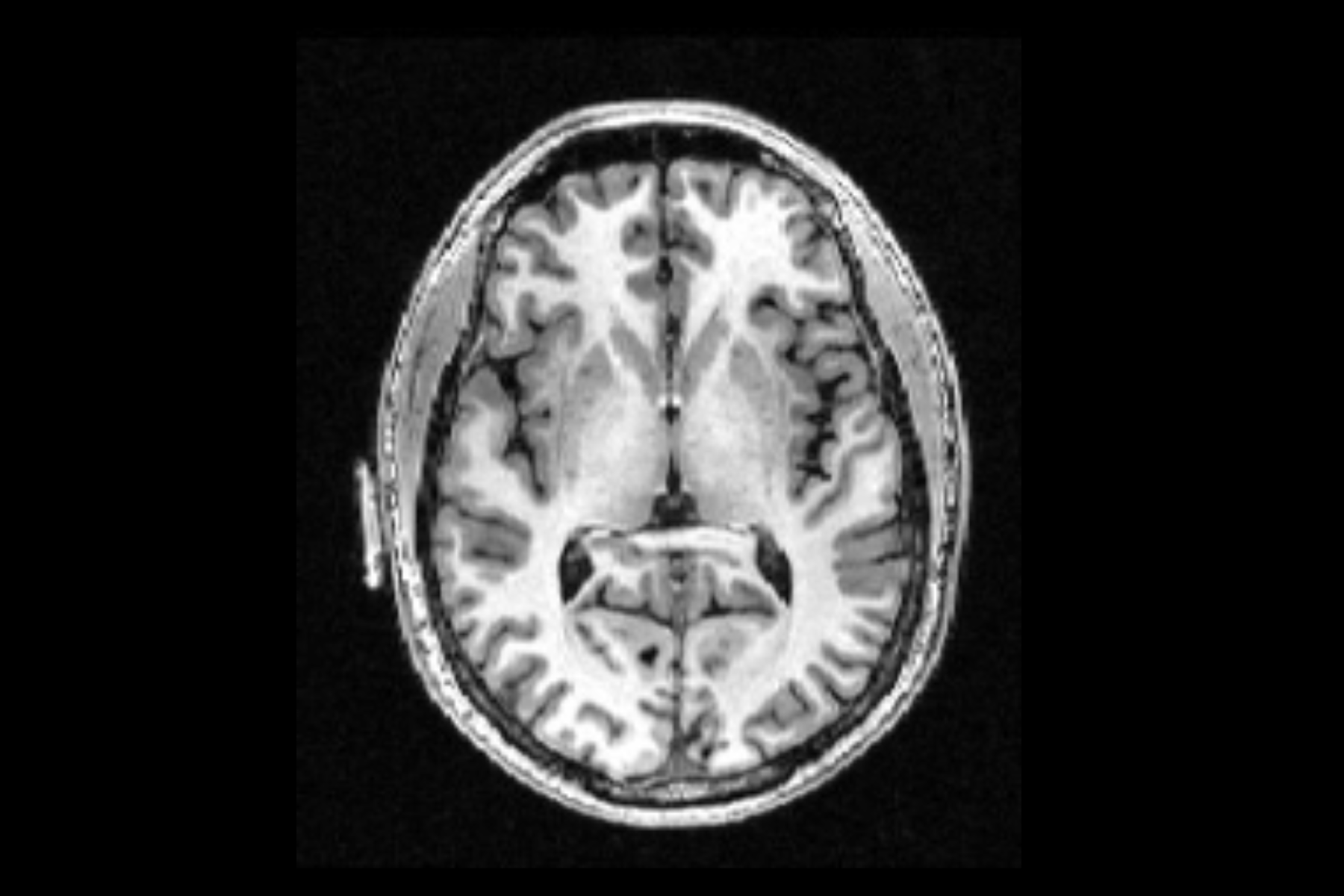
Mercadante AA, Tadi P. Neuroanatomy, Gray Matter. [Updated 2021 Jul 31]. In: StatPearls [Internet]. Treasure Island (FL): StatPearls Publishing; 2022 Jan-.

Schmahmann JD, Smith EE, Eichler FS, Filley CM. Cerebral white matter: neuroanatomy, clinical neurology, and neurobehavioral correlates. Ann N Y Acad Sci. 2008 Oct

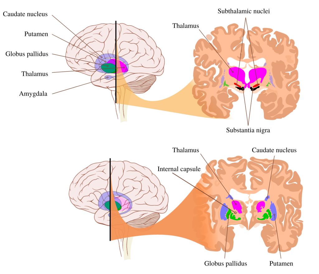
Young CB, Reddy V, Sonne J. Neuroanatomy, Basal Ganglia. [Updated 2021 Jul 31]. In: StatPearls [Internet]. Treasure Island (FL): StatPearls Publishing; 2022 Jan-.

Muzio MR, Cascella M. Histology, Axon. [Updated 2021 Nov 19]. In: StatPearls [Internet]. Treasure Island (FL): StatPearls Publishing; 2022 Jan-.

Azevedo FA, Carvalho LR, Grinberg LT, Farfel JM, Ferretti RE, Leite RE, Jacob Filho W, Lent R, Herculano-Houzel S. Equal numbers of neuronal and nonneuronal cells make the human brain an isometrically scaled-up primate brain. J Comp Neurol. 2009 Apr 10;513(5):532-41. doi: 10.1002/cne.21974. PMID: 19226510.

AbuHasan Q, Reddy V, Siddiqui W. Neuroanatomy, Amygdala. 2022 Jul 19. In: StatPearls [Internet]. Treasure Island (FL): StatPearls Publishing; 2022 Jan–.

Sonne J, Reddy V, Beato MR. Neuroanatomy, Substantia Nigra. 2021 Oct 30. In: StatPearls [Internet]. Treasure Island (FL): StatPearls Publishing; 2022 Jan–.

Further readings:
, ,