Isn’t it nice to hear birds singing, the voice of a loved one or our favourite song on the radio? All this is possible thanks to our hearing system!
*Text 1 of 4 in the series about speech sounds processing.
To understand how sound spreads from our ears to our brain, we propose to take you on a small tour of the auditory system, starting with the peripheral auditory system.
From a physical standpoint, sound is a pressure variation, caused by a vibrating object (e.g., vocal cords), that propagates in an elastic medium (e.g., air). These pressure variations cause the air molecules to move, and this movement is captured by the peripheral auditory system.
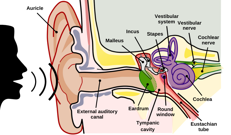
The peripheral auditory system is divided into three components: the outer ear, the middle ear and the inner ear (Figure 1).
The outer ear is made up of the auricle and the external auditory canal (see Figure 1). The auricle acts as a funnel and captures the sounds of the environment. Its shape helps locate the source of the sounds and amplifies the sounds of human speech. The outer ear also plays a protective role against certain foreign objects: the cerumen (ear wax) and the hairs of the external auditory canal make it possible to collect dust, bacteria, etc.
The middle ear is composed of the eardrum, the three ossicles (the malleus, the incus and the stapes), which are the smallest bones of the human body, and the Eustachian tube. The eardrum is a membrane. In the presence of pressure variations in the air (sound), the eardrum vibrates. This vibration induces a movement of the malleus, whose handle is embedded in the membrane of the eardrum. The vibration of the eardrum also causes the movement of the incus and stapes, the three ossicles being connected and forming what is called the chain of ossicles. The middle ear transmits the sound mechanically as the air pressure variations are transformed into a sequence of movements. In case of loud sounds (e.g., dog bark, car horn, MRI noise), muscles attached to the malleus and stapes contract automatically, reducing the mobility of the ossicle chain and limiting sound propagation. This reflex allows a certain control over the intensity of the sound meant to protect the inner ear.
For the eardrum to vibrate properly, the pressure must be balanced on both sides of the membrane. This is where the Eustachian tube, which connects the middle ear to the nasopharynx (upper part of the throat, behind the nose) intervenes. The Eustachian tube provides the middle ear with air and ensures that the pressure in the tympanic cavity is similar to the one in the external auditory canal.
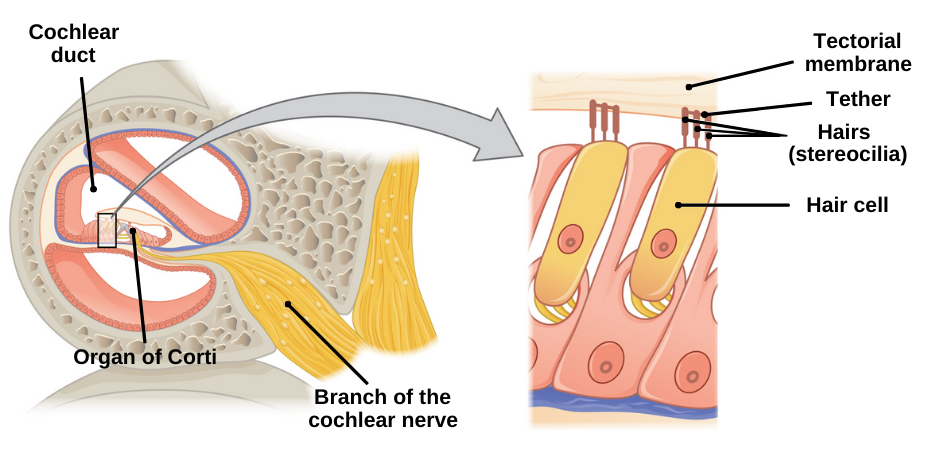
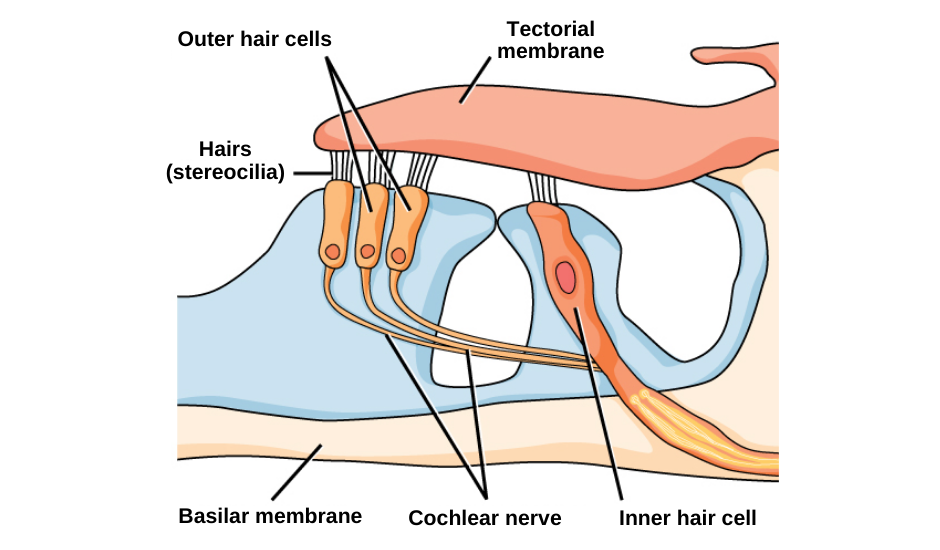
The inner ear is made up of the cochlea and the vestibular system, the organ responsible for balance. The cochlea is a small but complex organ. It is shaped as a spiral, with a length of approximately 35 mm, dug inside the temporal bone (a skull bone located near the temples). Inside the cochlea is the cochlear duct. This duct contains a fluid called endolymph, as well as the organ of Corti (see Figure 2). The organ of Corti contains the sensory cells that allow us to hear: the hair cells. These cells are called hair cells, because they have small hairs, called stereocilia. There are two types of hair cells: the inner and the outer hair cells (see Figure 3). The inner hair cells are the actual sensory receptors, while the outer hair cells serve to amplify to movement of the basilar membrane and increase its resolution.
Figure 3. Illustration of two types of hair cells: the inner and the outer hair cells (Biology) adapted from CNX OpenStax under licence CC BY 4.0.
The entrance to the cochlea is the oval window, to which the stapes is attached. When the ossicle chain is in motion, the stapes makes a piston movement, thus stirring the liquid contained in the cochlea. This movement causes the displacement of the membranes surrounding the cochlear duct and, in turn, the stereocilia are bent. The hair cells then send an electrical impulse, which is routed through the branches of the cochlear nerve to the brain. How does the brain know the intensity (strong or weak) of a sound? It works a bit like the Morse code! The sound intensity is encoded by the frequency of the pulses emitted by the hair cells. Stronger sounds result in greater activity (closer pulses) than weaker sounds. And what about the frequency of the sounds (low or high pitch)? The message sent to the brain depends on the place of the cochlea that enters in vibration. The high-pitched sounds make the base of the cochlea vibrate while the low-pitched sounds activate the top of the cochlea. This organization is called “tonotopy”.
This concludes the beginning of our journey into the auditory system. But this is only the beginning of the journey because the sounds must travel to the brain to be processed! Stay tuned for the upcoming texts in this series.