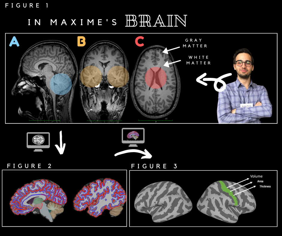
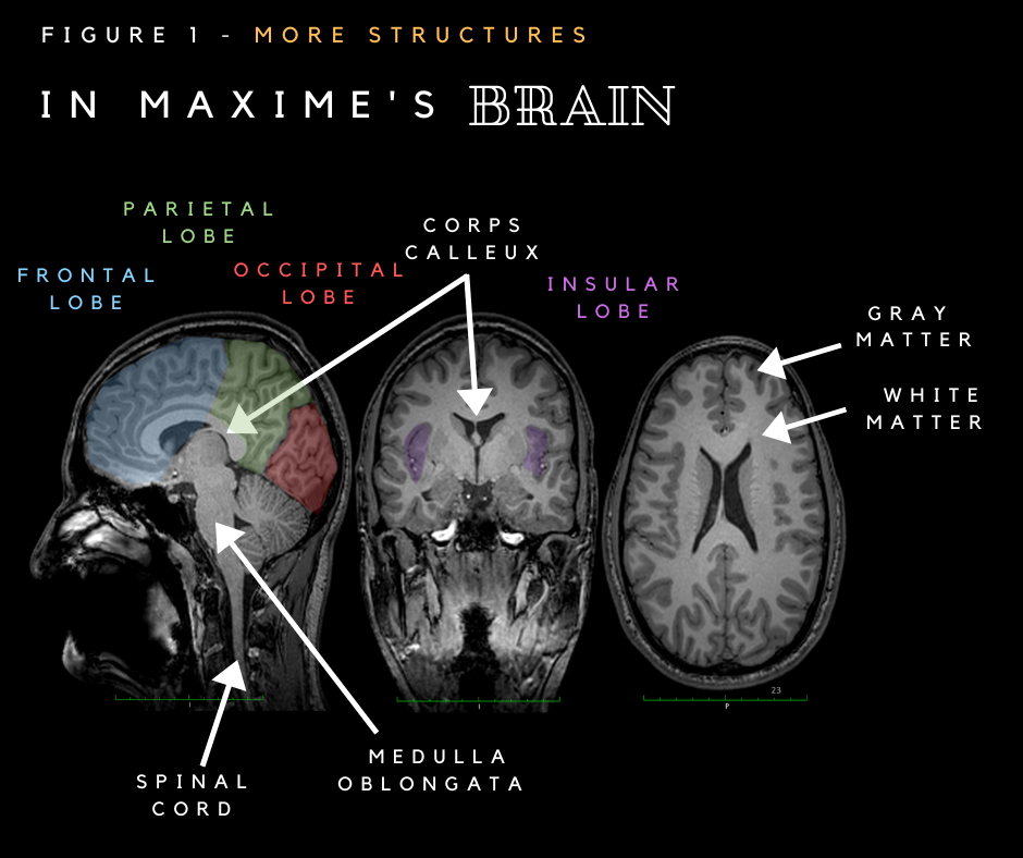
The anatomical images obtained by MRI illustrate on a gray scale the time taken by the hydrogen atoms, contained mainly in the molecules of water (H2O), to go back to their original position in space after having been spun by radio waves. (-> How it works – MRI blog article) It is possible to differentiate the structures and tissues of our body according to their water composition because this duration varies according to the composition of the tissues, which will be more or less dark on the images. In figure 1, we can see the difference in tones between the gray matter, containing the cell bodies of the neurons (in dark gray on the image) and the white matter, which contains their extensions, which can be conceptualized as electric wires (in pale gray on the image).
In A, we see the cerebellum, in B the temporal lobes and in C the cerebral ventricles.
In the lab, we use these images to study the anatomy of brain regions and examine the relationship between the anatomy of these regions and language and cognitive functions. We also seek to understand how aging and certain forms of activity can modify the anatomy of the brain and its functioning. Finally, we also use these images to target regions of the brain and then stimulate them using transcranial magnetic stimulation (TMS).
In order to study the structure of the gray matter on the surface of the cerebral cortex, the MRI images are processed in several stages with different specialized software in order to isolate the brain from the skull and, in a second step, to separate the gray from the white matter (Figure 2). In the figure, the contour of the gray matter is represented as a red line while the white matter contour is represented as a blue line. Like our fingerprints, the folds of our brain differ quite a bit between each person. The software is then used to smooth the brain (Figure 3), making the sulci and cerebral convolutions (also called gyrus), which form a series of sinuous folds of the cerebral cortex, easier to distinguish. It is then possible to measure the area, volume and thickness of regions of interest and to compare these measurements between groups of participants, e.g., young and old, musicians and non-musicians, bilingual and monolingual people, etc. This is what Maxime did in his master’s project.
We conclude that Maxime has a magnificent brain, and we thank him warmly for letting us use it for this article!